
New Advances in the Study of Doping Modification for NASICON-Type Solid Electrolytes
Recently, the Advanced Laser and Optoelectronic Functional Materials Department of the Shanghai Institute of Optics and Fine Mechanics, Chinese Academy of Sciences, has made significant progress in the study of doping modification of NASICON-type solid-state electrolytes. The team revealed the mechanism by which La doping regulates the microstructure and ionic conductivity of NASICON-type Na3+xLaxZr2-xSi2PO12 solid-state electrolytes. The related research results have been published in The Journal of Physical Chemistry C under the title “The Impact of La Doping on the Ionic Conductivity of Na3+xLaxZr2-xSi2PO12 Solid-State Electrolytes.”
NASICON-type solid-state electrolytes hold great promise for applications in electrochemical energy storage, with improving their ionic conductivity being a major research focus. Previous studies have demonstrated that substituting Zr4+ with divalent or trivalent cations in Na3Zr2Si2PO12 can significantly enhance ionic conductivity. However, the underlying mechanism remains under debate—whether the improvement stems from compositional changes or increased material densification has not been definitively resolved.
In this study, advanced solid-state nuclear magnetic resonance (NMR) techniques were employed to systematically investigate the influence of La3+ doping on the structure, morphology, and electrochemical performance of the material. The results revealed that La3+ primarily segregates at the grain boundaries to form a Na3La(PO4)2 secondary phase, rather than substituting Zr4+ within the main phase. Although La doping alters the Si/P ratio and Na+ concentration in the primary phase and induces a phase transition, experimental evidence confirms that the key factor driving the enhancement in ionic conductivity is the improved material densification.
This finding provides new theoretical insights into the optimization of NASICON-type solid-state electrolytes.
This work was supported by the Strategic Priority Research Program of the Chinese Academy of Sciences (XDB0650000).
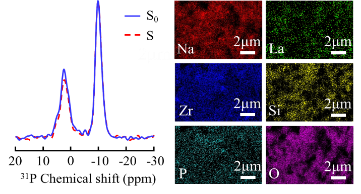
Left: Comparison of the signal intensity (S0) without π pulse applied to 139La nucleus and signal intensity (S) when π pulse is reapplied to 139La nucleus of the sample with x = 0.4 at a fixed time of NTr = 1.15 ms. Right: Elemental mapping images of the x = 0.4 sample for Na, La, Zr, Si, P, and O
Article link: https://doi.org/10.1021/acs.jpcc.4c07861
Contact: REN Jinjun
Advanced Laser and Optoelectronic Functional Materials Department,
Shanghai Institute of Optics and Fine Mechanics, CAS
Email: jinjunren@siom.ac.cn@siom.ac.cn